Search Thermo Fisher Scientific
Disclaimer

Clicking the images or links will redirect you to a website hosted by BenchSci that provides third-party scientific content. Neither the content nor the BenchSci technology and processes for selection have been evaluated by us; we are providing them as-is and without warranty of any kind, including for use or application of the Thermo Fisher Scientific products presented.
")
 in Flow")
 in Flow")
 in Flow")
 in Flow")
产品信息
50-0831-82
应用
建议稀释比
已发表文章
产品规格
种属反应
Mouse
已发表种属
Not Applicable
宿主/亚型
Rat
/ IgG1
分类
Monoclonal
类型
Antibody
克隆号
Michel-17 (Michel17)
偶联物
激发/发射光谱
651/668 nm
查看光谱
形式
Liquid
浓度
0.2 mg/mL
纯化类型
Affinity chromatography
保存液
PBS, pH 7.2
内含物
0.09% sodium azide
保存条件
4° C, store in dark, DO NOT FREEZE!
运输条件
Ambient (domestic); Wet ice (international)
RRID
AB_10609344
产品详细信息
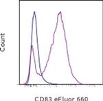
Description: The Michel-17 monoclonal antibody reacts with mouse CD83, a 45kDa cell surface glycoprotein and a member of the Ig superfamily. The mouse CD83 antigen is expressed predominantly on mature DC and activated lymphocytes. Cross-linking of CD83 with Michel-17 on DC or activated T cells does not induce any activation signal. CD83 plays an important role in T cell development through interaction with its ligand. CD83-Ig protein has revealed the presence of a CD83 ligand expressed mainly by B220+ cells in mouse spleen.
Applications Reported: This Michel-17 (Michel17) antibody has been reported for use in flow cytometric analysis.
Applications Tested: This Michel-17 (Michel17) antibody has been tested by flow cytometric analysis of stimulated mouse splenocytes. This can be used at less than or equal to 0.25 µg per test. A test is defined as the amount (µg) of antibody that will stain a cell sample in a final volume of 100 µL. Cell number should be determined empirically but can range from 10^5 to 10^8 cells/test. It is recommended that the antibody be carefully titrated for optimal performance in the assay of interest.
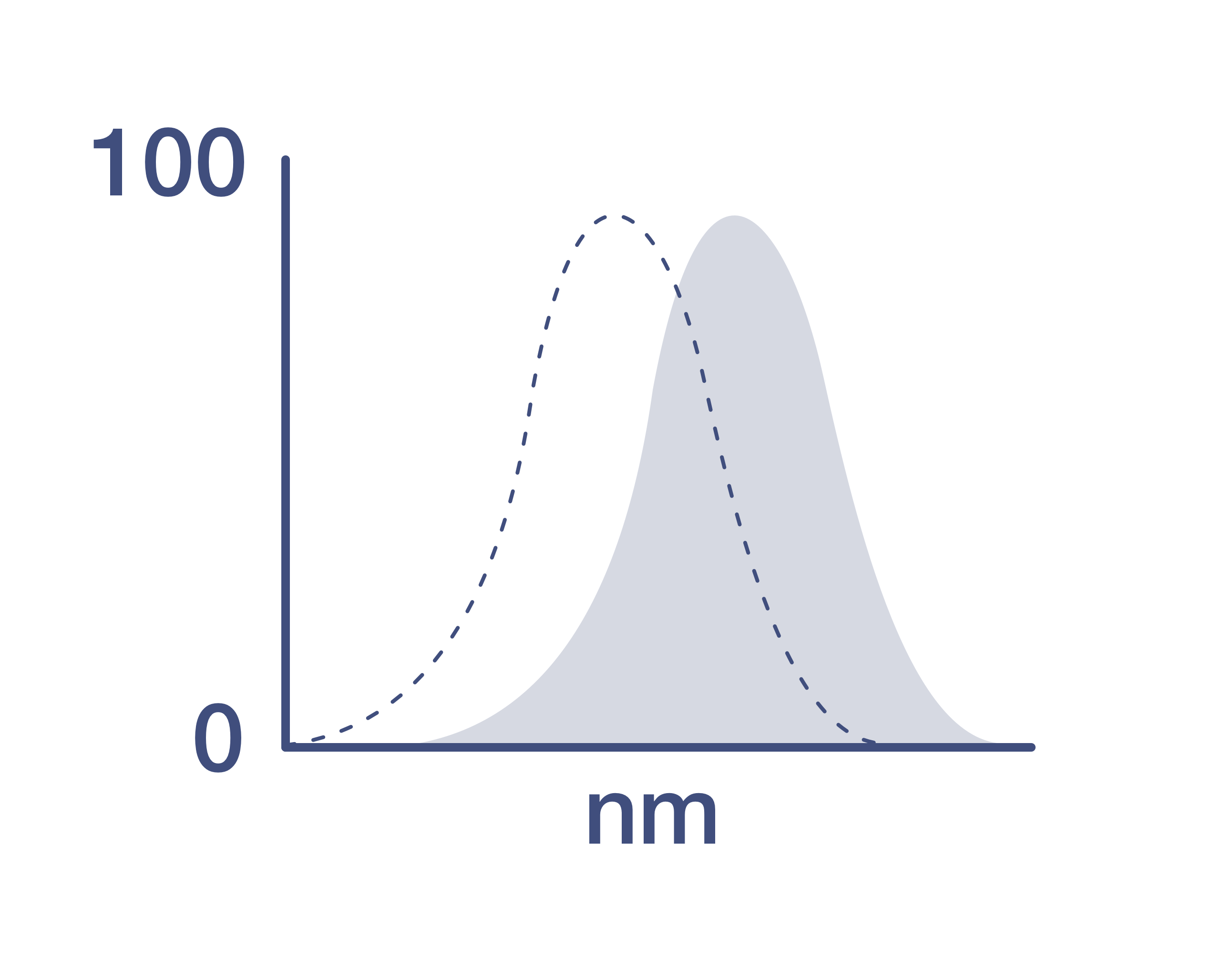
eFluor® 660 is a replacement for Alexa Fluor® 647. eFluor® 660 emits at 659 nm and is excited with the red laser (633 nm). Please make sure that your instrument is capable of detecting this fluorochome.
Excitation: 633-647 nm; Emission: 668 nm; Laser: Red Laser.
Filtration: 0.2 µm post-manufacturing filtered.
靶标信息
CD83 cell surface antigen is a 40-45kD glycoprotein expressed by peripheral blood dendritic cells. Peripheral lymphocytes can be induced to express very low levels of CD83 after culture in agents such as Con A or PHA. In immunohistology, CD83 is shown to be expressed strongly by interfollicular interdigitating reticulum cells and more weakly by cells within germinal centres. CD83 is also expressed by Langerhan's cells in the skin. The CD83 antigen is a 186-amino-acid single-chain glycoprotein and this molecule is a member of the immunoglobulin superfamily that is composed of an extracellular V-type Ig-like single domain, a transmembrane region, and a short, 40-amino-acid cytoplasmic tail. CD83 antigen undergoes extensive post-translational glycosylation, since the determined Mr is twice the predicted size of the core protein. However, CD83+ cells have a unique cell surface immuno-phenotype that does not correlate with that of T cells, B cells, NK cells, or cells of the myelomonocytic lineage. CD83+ cells coexpress the highest levels of MHC class II molecules, when compared with other leucocyte lineages. They also co-express T cell markers (CD2, CD5), B cell markers (CD40, CD78), myeloid cell markers (CD13, CD33, CD36), cytokine receptors as well as other cell surface molecules. Diseases associated with CD83 dysfunction include plague and Rift Valley Fever.
仅用于科研。不用于诊断过程。未经明确授权不得转售。
How to use the Panel Builder
Watch the video to learn how to use the Invitrogen Flow Cytometry Panel Builder to build your next flow cytometry panel in 5 easy steps.
生物信息学
蛋白别名: CD antigen CD83; CD83; CD83 antigen; mCD83
基因别名: Cd83
UniProt ID: (Mouse) O88324
Entrez Gene ID: (Mouse) 12522

