Search Thermo Fisher Scientific
Disclaimer

Clicking the images or links will redirect you to a website hosted by BenchSci that provides third-party scientific content. Neither the content nor the BenchSci technology and processes for selection have been evaluated by us; we are providing them as-is and without warranty of any kind, including for use or application of the Thermo Fisher Scientific products presented.
 Antibody in Immunocytochemistry (ICC/IF)")
图: 1 / 5
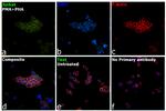
CD279 (PD-1) Antibody (14-2798-82) in ICC/IF
Immunofluorescence analysis of Programmed cell death protein 1 was performed using 70% confluent log phase Jurkat cells treated with PMA and PHA (50 ng/mL PHA and 1 mg/mL PMA, for 48 hours). The cells were fixed with 4% paraformaldehyde for 10 minutes, permeabilized with 0.1% Triton™ X-100 for 15 minutes, and blocked with 2% BSA for 45 minutes at room temperature. The cells were labeled with CD279 (PD-1) Mo... View More


 Antibody (14-2798-82) in ICC/IF")
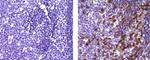
 Antibody (14-2798-82) in IHC (P)")
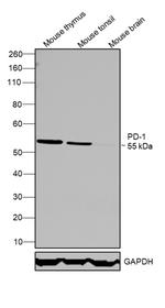
 Antibody (14-2798-82) in WB")
 Antibody (14-2798-82)")
 Antibody (14-2798-82)")
产品信息
14-2798-82
产品规格
种属反应
Human,
Mouse
宿主/亚型
Mouse
/ IgG1, kappa
分类
Monoclonal
类型
Antibody
克隆号
J121
偶联物
形式
Liquid
浓度
0.5 mg/mL
纯化类型
Affinity chromatography
保存液
PBS, pH 7.2
内含物
0.09% sodium azide
保存条件
4° C
运输条件
Ambient (domestic); Wet ice (international)
RRID
AB_2572870
产品详细信息
Description: This J121 monoclonal antibody reacts with human CD279 (programmed death-1, PD-1), a 55 kDa member of the CD28 immunoglobulin superfamily. CD279 contains the immunoreceptor tyrosine-based inhibitory motif (ITIM) and plays a key role in peripheral tolerance and autoimmune disease. CD279 is expressed predominantly on activated T and B lymphocytes. Two novel members of the B7 family have been identified as the CD279 ligands, CD274 (PD-L1, B7-H1) and CD273 (PD-L2, B7-DC). Evidence reported to date suggests overlapping functions for these two ligands and their constitutive expression on some normal tissues and upregulation on activated antigen-presenting cells. More recently, therapies targeting the blockade of the CD279/CD274 pathway have become the focus for treatment of melanoma, renal cell cancer, Hodgkins' lymphoma, and non-small cell lung carcinoma (NSCLC).
The J121 monoclonal antibody is not recommended for flow cytometry of human cells. For detection of human CD279 using flow cytometry please refer to clone eBioJ105 (J105).
Applications Reported: This J121 antibody has been reported for use in western blotting, and immunohistochemical staining of formalin-fixed paraffin embedded tissue sections.
Applications Tested: This J121 antibody has been tested by immunohistochemistry of formalin-fixed paraffin embedded human tissue using high or low pH antigen retrieval and can be used at less than or equal to 5 µg/mL. It is recommended that the antibody be carefully titrated for optimal performance in the assay of interest.
Purity: Greater than 90%, as determined by SDS-PAGE.
Aggregation: Less than 10%, as determined by HPLC.
Filtration: 0.2 µm post-manufacturing filtered.
靶标信息
Cell-mediated immune responses are initiated by T lymphocytes that are themselves stimulated by cognate peptides bound to MHC molecules on antig en-presenting cells (APC). T-cell activation is generally self-limited as activated T cells express receptors such as PD-1 (also known as PDCD-1) that mediate inhibitory signals from the APC. PD-1 can bind two different but related ligands, PDL-1 and PDL-2. Upon binding to either of these ligands, signals generated by PD-1 inhibit the activation of the immune response in the absence of "danger signals" such as LPS or other molecules associated with bacteria or other pathogens. Evidence for this is seen in PD1-null mice who exhibit hyperactivated immune systems and autoimmune diseases. Despite its predicted molecular weight, PD-1 often migrates at higher molecular weight in SDS-PAGE.
仅用于科研。不用于诊断过程。未经明确授权不得转售。
篇参考文献 (0)
您是否在文献中引用过该产品?请点击下方按钮邮件告知我们。
生物信息学
蛋白别名: CD279; hPD1; mPD-1; programmed cell death 1 protein; Programmed cell death protein 1; Protein PD-1; Protein PD1; sCD279; soluble CD279; systemic lupus erythematosus susceptibility 2
基因别名: CD279; hPD-1; hPD-l; hSLE1; Ly101; PD-1; PD1; Pdc1; PDCD1; SLEB2
UniProt ID: (Human) Q15116, (Mouse) Q02242
Entrez Gene ID: (Human) 5133, (Mouse) 18566
negative regulation of tolerance induction
apoptotic process
humoral immune response
signal transduction
multicellular organism development
T cell costimulation
negative regulation of apoptotic process
positive regulation of T cell apoptotic process
immune system process
positive regulation of apoptotic process

