Search Thermo Fisher Scientific
Disclaimer

Clicking the images or links will redirect you to a website hosted by BenchSci that provides third-party scientific content. Neither the content nor the BenchSci technology and processes for selection have been evaluated by us; we are providing them as-is and without warranty of any kind, including for use or application of the Thermo Fisher Scientific products presented.
")
图: 1 / 5
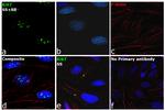
Ki-67 Antibody (PA1-38032) in ICC/IF
Immunofluorescence analysis of Ki67 was performed using 70% confluent log phase HeLa cells serum starved for 36 hours followed by serum release for 6 hrs. The cells were fixed with 4% Paraformaldehyde for 10 minutes, permeabilized with 0.1% Triton™ X-100 for 10 minutes, and blocked with 2% BSA for 10 minutes at room temperature. The cells were labeled with Ki67 Polyclonal Antibody (Product # PA1-38032) at 1:100 dilution in 0.1% BSA, incubated at 4 degree Celsius overnight and then labeled with Goat anti-Rabbit IgG (Heavy Chain) Superclonal... View More

 in ICC/IF")
 in IHC (P)")
 in IHC")
")
产品信息
PA1-38032
应用
建议稀释比
已发表文章
产品规格
种属反应
Human
已发表种属
Human,
Mouse
宿主/亚型
Rabbit
/ IgG
分类
Polyclonal
类型
Antibody
抗原
Synthetic peptide derived from C-terminus of human Ki-67 protein
偶联物
形式
Liquid
浓度
0.12 mg/mL
纯化类型
Antigen affinity chromatography
保存液
TBS, pH 7.6, with 1% BSA
内含物
0.1% sodium azide
保存条件
Store at 4°C short term. For long term storage, store at -20°C, avoiding freeze/thaw cycles.
运输条件
Gel ice packs
RRID
AB_2142239
产品详细信息
This antibody is predicted to react with bovine based on sequence homology.
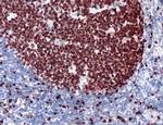
Heat-mediated antigen retrieval is recommended prior to staining, using a 10mM citrate buffer, pH 6.0, for 10 minutes followed by cooling at room temperature for 20 min. Following antigen retrieval, incubate samples with primary antibody for 10 min at room temperature. A suggested positive control is tonsil, lymph node or breast carcinoma.
靶标信息
Ki-67 is a nuclear protein that is expressed during various stages in the cell cycle, particularly during late G1, S, G2, and M phases. The protein has a forkhead associated domain (FHA) through which it associates with euchromatin at the perichromosomal layer, the centromeric heterochromatin, and the nucleolus. Ki-67 is shown to have a cell cycle dependent topographical distribution with perinucleolar expression at G1, expression in the nuclear matrix at G2, and expression on the chromosomes during M phase. Ki-67 is commonly used as a proliferation marker because it is not detected in G0 cells, but increases steadily from G1 through mitosis. Ki-67 antibodies are useful in establishing the cell growing fraction in neoplasms. In neoplastic tissues, the prognostic value is comparable to the tritiated thymidine-labelling index. The correlation between low Ki-67 index and histologically low-grade tumors is strong. Ki-67 is routinely used as a neuronal marker of cell cycling and proliferation.
仅用于科研。不用于诊断过程。未经明确授权不得转售。
生物信息学
蛋白别名: Antigen identified by monoclonal antibody Ki-67; Antigen KI-67; Proliferation marker protein Ki-67; proliferation-related Ki-67 antigen; protein phosphatase 1, regulatory subunit 105; RP11-380J17.2
基因别名: KIA; MIB-; MIB-1; MKI67; PPP1R105
UniProt ID: (Human) P46013
Entrez Gene ID: (Human) 4288

