Search Thermo Fisher Scientific
Disclaimer

Clicking the images or links will redirect you to a website hosted by BenchSci that provides third-party scientific content. Neither the content nor the BenchSci technology and processes for selection have been evaluated by us; we are providing them as-is and without warranty of any kind, including for use or application of the Thermo Fisher Scientific products presented.
")
图: 1 / 1
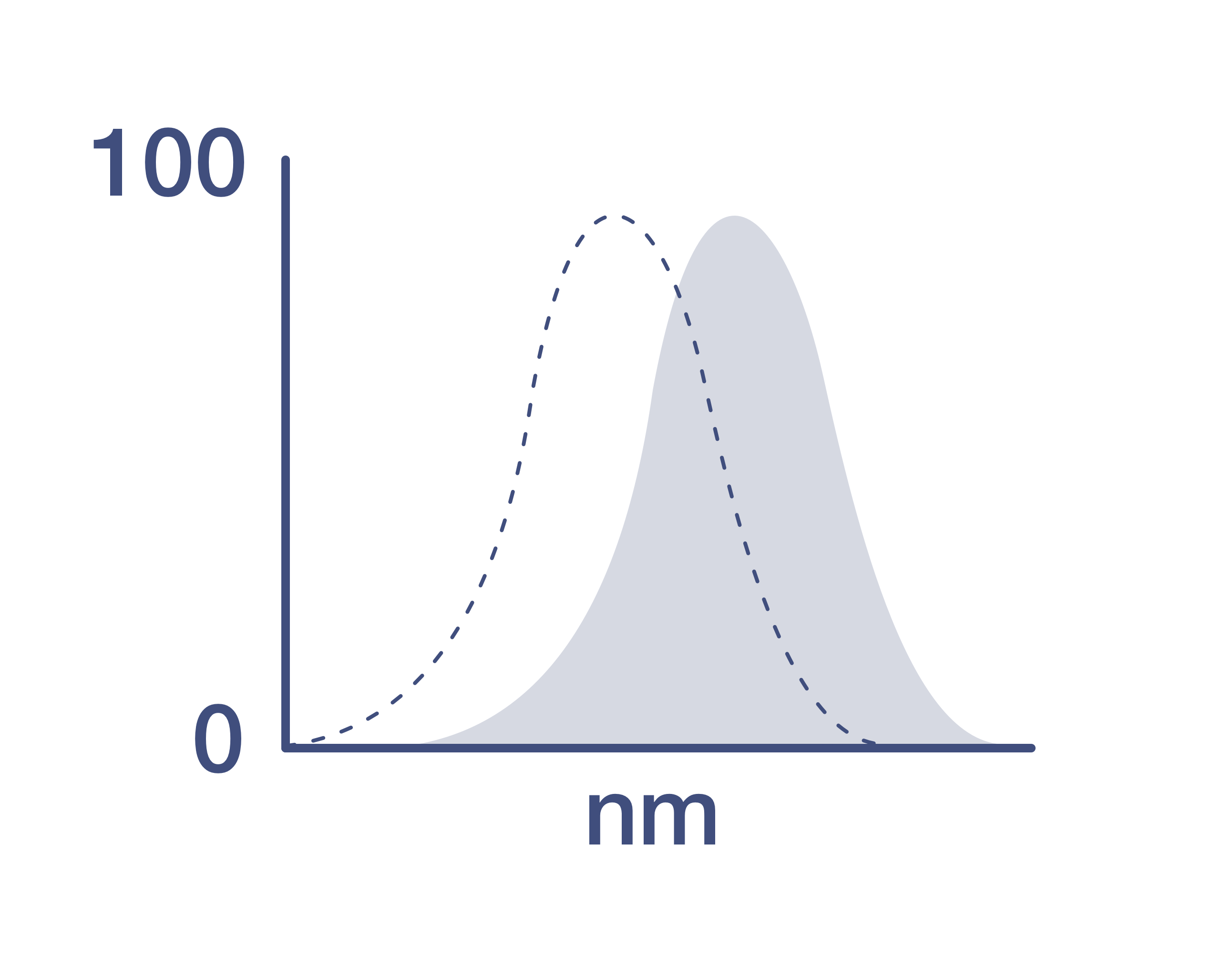
MCL-1 Antibody (12-9047-42) in Flow
Normal human peripheral blood cells were untreated (blue histogram) or stimulated for 72 hours with CD3 and CD28 Monoclonal Antibodies, Functional Grade (Product # 16-0037-85 and Product # 16-0289-85) and MG-132 (purple histogram). Cells were then stained intracellularly, using the Intracellular Fixation & Permeabilization Buffer Set (Product # 88-8824-00) and protocol, with MCL-1 Monoclonal Antibody, PE. Cells in the lymphocyte gate were used for analysis.

 in Flow")
产品信息
12-9047-42
应用
建议稀释比
已发表文章
产品规格
种属反应
Human,
Mouse
已发表种属
Mouse
宿主/亚型
Mouse
/ IgG1, kappa
分类
Monoclonal
类型
Antibody
克隆号
LVUBKM
偶联物
激发/发射光谱
565/576 nm
查看光谱
形式
Liquid
浓度
5 µL/Test
纯化类型
Affinity chromatography
保存液
PBS, pH 7.2, with 0.2% BSA
内含物
0.09% sodium azide
保存条件
4° C, store in dark, DO NOT FREEZE!
运输条件
Ambient (domestic); Wet ice (international)
RRID
AB_2762598
产品详细信息
Description: This LVUBKM monoclonal antibody recognizes human and mouse myeloid cell leukemia sequence 1 (Mcl-1). Mcl-1 is an anti-apoptotic member of the Bcl-2 family of proteins important for regulation of cell survival/apoptosis. Mcl-1 is primarily localized to the outer membrane of mitochondria where it prevents cytochrome c release via dimerization with other Bcl-2 family members such as Bim. Although it is expressed in both immune and non-immune cells, highest levels of Mcl-1 expression are seen in hematopoietic lineage cells. PI3K activation of AKT results in destabilization and degradation of GSK3 beta, which prevents phosphorylation of Mcl-1 on S159 and its subsequent ubiquitnation and degradation. Mice conditionally lacking Mcl-1 in lymphocytes showed that Mcl-1 is essential during early lymphoid development and for the maintenance of mature lymphocytes.
Applications Reported: This LVUBKM antibody has been reported for use in flow cytometric analysis.
Applications Tested: This LVUBKM antibody has been pre-diluted and tested by flow cytometric analysis of stimulated normal human peripheral blood cells. This may be used at 5 µL (0.25 µg) per test. A test is defined as the amount (µg) of antibody that will stain a cell sample in a final volume of 100 µL. Cell number should be determined empirically but can range from 10^5 to 10^8 cells/test.
Excitation: 488-561 nm; Emission: 578 nm; Laser: Blue Laser, Green Laser, Yellow-Green Laser
靶标信息
MCL1 (Myeloid cell leukemia-1) belongs to the Bcl-2 family and is involved in programing, differentiation and concomitant maintenance of cell viability, but not of proliferation. Isoform 1 of MCL1 inhibits apoptosis while isoform 2 promotes it. The carboxy terminal of MCL1 and bcl-2 share significant sequence homology. Expression of MCL1 is increased upon exposure of ML-1 cells to various types of DNA damaging agents (e.g. ionizing radiation, ultraviolet radiation, and alkylating drugs) along with increases in GADD45 and Bax and a decrease in bcl-2. Enhanced expression of MCL1, prominently associated with mitochondria, complements the continued expression of bcl-2 in ML-1 cells undergoing differentiation. Like bcl-2, MCL1 has the capacity to promote cell viability under conditions that otherwise cause apoptosis. While the mechanism by which MCL1 inhibits apoptosis is not known, it is thought that it may heterodimerize and neutralize pro-apoptotic members of the Bcl-2 family such as Bim or Bak. MCL1 was originally identified in differentiating myeloid cells, but has since been shown to be expressed in multiple cell types. MCL1 is essential for embryogenesis and for the development and maintenance of B and T lymphocytes in animals. MCL1 exists as at least two distinct isoforms designated MCL1L and MCL1S. In marked contrast to the larger isoform of MCL1, overexpression of MCL1S promotes cell death.
仅用于科研。不用于诊断过程。未经明确授权不得转售。
How to use the Panel Builder
Watch the video to learn how to use the Invitrogen Flow Cytometry Panel Builder to build your next flow cytometry panel in 5 easy steps.
生物信息学
蛋白别名: Bcl-2-like protein 3; Bcl-2-related protein EAT/mcl1; Bcl2-L-3; Induced myeloid leukemia cell differentiation protein Mcl-1; Induced myeloid leukemia cell differentiation protein Mcl-1 homolog; MCL-1S; mcl1/EAT; MGC104264; MGC1839; myeloid cell leukemia 1; myeloid cell leukemia ES; myeloid cell leukemia sequence 1 (BCL2-related)
基因别名: AW556805; bcl2-L-3; BCL2L3; EAT; Mcl-1; MCL1; MCL1-ES; mcl1/EAT; MCL1L; MCL1S; TM
UniProt ID: (Human) Q07820, (Mouse) P97287
Entrez Gene ID: (Human) 4170, (Mouse) 17210
cell fate determination
multicellular organism development
intrinsic apoptotic signaling pathway in response to DNA damage
apoptotic mitochondrial changes
cellular homeostasis
response to cytokine
negative regulation of apoptotic process
protein transmembrane transport
extrinsic apoptotic signaling pathway in absence of ligand
positive regulation of oxidative stress-induced neuron intrinsic apoptotic signaling pathway
negative regulation of anoikis
regulation of response to DNA damage stimulus
negative regulation of extrinsic apoptotic signaling pathway in absence of ligand
negative regulation of intrinsic apoptotic signaling pathway
apoptotic process
cell differentiation
regulation of apoptotic process

