Search Thermo Fisher Scientific
Disclaimer

Clicking the images or links will redirect you to a website hosted by BenchSci that provides third-party scientific content. Neither the content nor the BenchSci technology and processes for selection have been evaluated by us; we are providing them as-is and without warranty of any kind, including for use or application of the Thermo Fisher Scientific products presented.
 Antibody in Immunocytochemistry (ICC/IF)")
图: 1 / 8
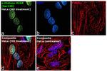
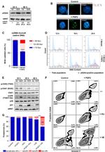
Phospho-Histone H2A.X (Ser139) Antibody (53-9865-82) in ICC/IF
Immunofluorescence analysis of phospho-H2A.X was performed using 70% confluent log phase HeLa cells treated with hydroxyurea (3 mM for 6 hr). The cells were fixed with 4% paraformaldehyde for 10 minutes, permeabilized with 0.1% Triton™ X-100 for 10 minutes, and blocked with 2% BSA for 45 minutes at room temperature. The cells were labeled with Phospho-Histone H2A.X (Ser139) Monoclonal Antibody (CR55T33), Al... View More

 Antibody (53-9865-82) in ICC/IF")
 Antibody (53-9865-82) in IHC (P)")
 Antibody (53-9865-82) in WB")
 Antibody (53-9865-82) in IHC")
 Antibody (53-9865-82) in WB")
 Antibody (53-9865-82) in WB")
 Antibody (53-9865-82) in Flow")
 Antibody (53-9865-82)")
产品信息
53-9865-82
应用
建议稀释比
已发表文章
产品规格
种属反应
Human,
Mouse
已发表种属
Human
宿主/亚型
Mouse
/ IgG1, kappa
分类
Monoclonal
类型
Antibody
克隆号
CR55T33
偶联物
激发/发射光谱
499/520 nm
查看光谱
形式
Liquid
浓度
0.5 mg/mL
纯化类型
Affinity chromatography
保存液
PBS, pH 7.2
内含物
0.09% sodium azide
保存条件
4° C, store in dark, DO NOT FREEZE!
运输条件
Ambient (domestic); Wet ice (international)
RRID
AB_2574485
产品详细信息
Description: The CR55T33 monoclonal antibody recognizes phosphorylated serine 139 of human and mouse H2AX. H2AX is a member of the H2A histone family that complex with DNA and other histones to form the repeating nucleosome units characteristic of eukaryotic chromatin. Nucleosomes consist of approximately 147 base pairs of DNA wrapped around an octamer of histones composed of two each of the four histone proteins: H2A, H2B, H3 and H4. After induction of DNA damage such as double-strand breaks by irradiation, genotoxic stresses, replication errors or gene recombination, PI3K-like kinases (e.g., ataxia telangiectasia mutated (ATM), ataxia telangiectasia Rad-3-related (ATR), and DNA-dependent protein kinase (DNA-PK) are activated to phosphorylate serine 139 in H2AX. This early phosphorylation event plays a critical role in recruiting proteins involved in DNA repair.
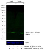
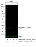
The monoclonal antibody CR55T33 recognizes a single band of approximately 15 kDa on reduced cell lysates from Jurkat cells stimulated with etoposide.
Applications Reported: This CR55T33 antibody has been reported for use in immunohistochemical staining of formalin-fixed paraffin embedded tissue sections, microscopy, and immunocytochemistry.
Applications Tested: This CR55T33 antibody has been tested by immunohistochemistry of formalin-fixed paraffin embedded human tissue using low or high pH antigen retrieval and can be used at less than or equal to 10 µg/mL. It is recommended that the antibody be carefully titrated for optimal performance in the assay of interest.
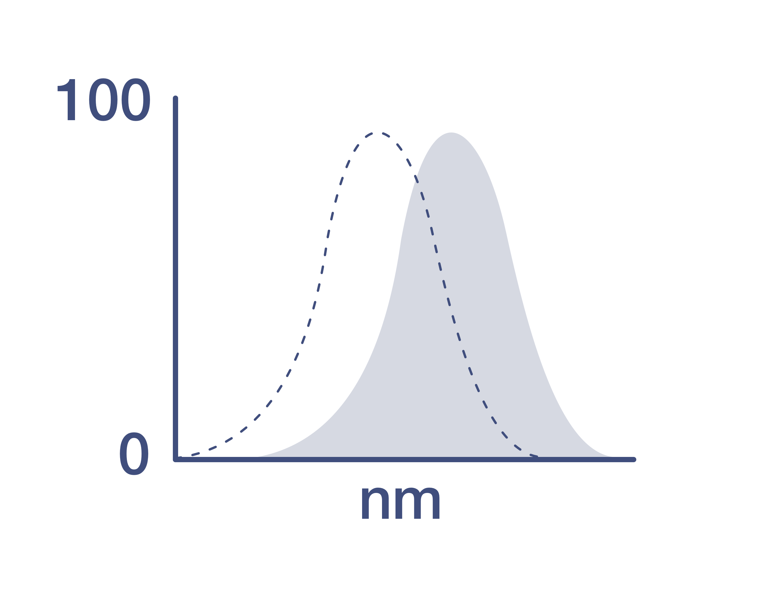
Excitation: 488 nm; Emission: 519 nm; Laser: Blue Laser.
Filtration: 0.2 µm post-manufacturing filtered.
靶标信息
Histone H2A.X (H2AX) is a member of the histone H2A family which is one of the four core histones making up the nucleosome core particle. In eukaryotes, DNA double strand breaks (DSBs) have been shown to trigger the phosphorylation of serine 139 at the carboxy terminus of histone H2AX resulting in gamma-H2AX. The phosphorylation of H2AX can be detected by Western blotting or immunofluorescence, revealing the frequency of DSBs. The phosphatidylinositol 3-kinases have been implicated in H2AX phosphorylation, but it is unclear if ATM is the primary H2AX kinase or if other members of the family such as DNA-PK and ATR contribute in a similar manner. Structurally, H2A.x contains 143 amino acid residues. Histone H2A.X is considered a basal histone, being synthesized in G1 as well as in S-phase, and its mRNA contains polyA addition motifs and a polyA tail along with the conserved stem-loop and U7 binding sequences involved in the processing and stability of replication type histone mRNAs. There are two forms of Histone H2A.X mRNA, one about 1600 bases long and contains polyA; the other about 575 bases long, lacking polyA. The short form behaves as a replication type histone mRNA, while the longer behaves as a basal type histone mRNA. Histone H2A.X maps to the 11q23.2-q23.3 region of the human chromosome. Histone H2A.x contributes to histone-formation and therefore the structure of DNA. Histone H2A variant H2A.x specifically regulates the interaction of MDC1 (mediator of DNA damage checkpoint protein 1), a DNA repair protein to the sites of DNA damage.
仅用于科研。不用于诊断过程。未经明确授权不得转售。
生物信息学
蛋白别名: gamma H2AX; H2A histone family, member X; H2a/x; H2AX histone; histone 5 protein 2ax; Histone H2A.X; Histone H2a/x; Histone H2AX
基因别名: AW228881; gammaH2ax; H2A.X; H2A/X; H2AFX; H2AX; Hist5-2ax
UniProt ID: (Human) P16104, (Mouse) P27661
Entrez Gene ID: (Human) 3014, (Mouse) 15270
DNA damage checkpoint
double-strand break repair via homologous recombination
double-strand break repair
double-strand break repair via nonhomologous end joining
nucleosome assembly
chromatin silencing
cellular response to DNA damage stimulus
spermatogenesis
response to ionizing radiation
viral process
cerebral cortex development
positive regulation of DNA repair
meiotic cell cycle
cellular response to gamma radiation
cellular senescence
DNA repair
DNA recombination
cell cycle

