Search Thermo Fisher Scientific
Disclaimer

Clicking the images or links will redirect you to a website hosted by BenchSci that provides third-party scientific content. Neither the content nor the BenchSci technology and processes for selection have been evaluated by us; we are providing them as-is and without warranty of any kind, including for use or application of the Thermo Fisher Scientific products presented.
")
图: 1 / 2
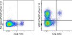
VISTA Antibody (46-1083-82) in Flow
Mouse splenocytes were stained with CD8a Monoclonal Antibody, FITC (Product # 11-0081-82) and 0.25 µg of Rat IgG2a kappa Isotype Control, PerCP-eFluor 710 (Product # 46-4321-82) (left) or 0.25 µg of VISTA Monoclonal Antibody, PerCP-eFluor 710 (right). Cells in the lymphocyte gate were used for analysis.
 in Flow")
")
产品信息
46-1083-82
产品规格
种属反应
Mouse
宿主/亚型
Rat
/ IgG2a, kappa
分类
Monoclonal
类型
Antibody
克隆号
MIH64
偶联物
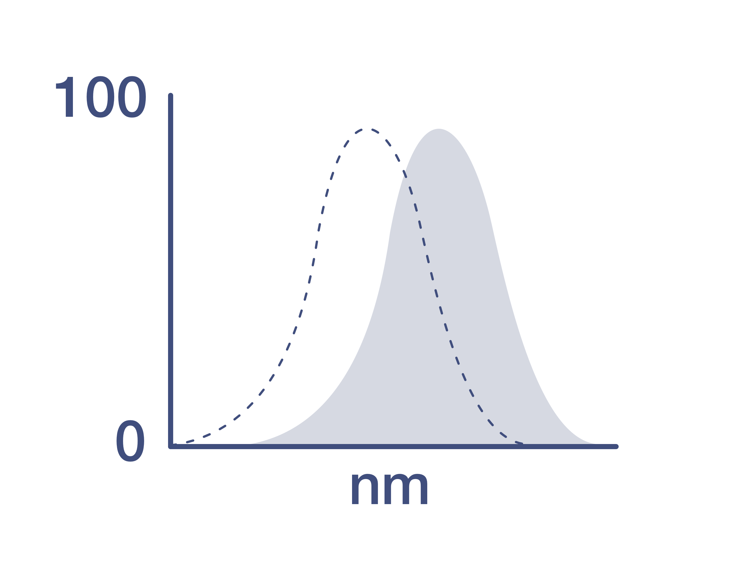
激发/发射光谱
482/708 nm
查看光谱
形式
Liquid
浓度
0.2 mg/mL
纯化类型
Affinity chromatography
保存液
PBS, pH 7.2
内含物
0.09% sodium azide
保存条件
4° C, store in dark, DO NOT FREEZE!
运输条件
Ambient (domestic); Wet ice (international)
RRID
AB_2725659
产品详细信息
Description: This MIH64 monoclonal antibody recognizes mouse VISTA also known as B7-H5 or PD-H1.
This MIH64 antibody will work in flow cytometry on both native and paraformaldehyde-fixed cells.
Applications Reported: This MIH64 antibody has been reported for use in flow cytometric analysis.
Applications Tested: This MIH64 antibody has been tested by flow cytometric analysis of mouse splenocytes. This may be used at less than or equal to 0.5 µg per test. A test is defined as the amount (µg) of antibody that will stain a cell sample in a final volume of 100 µL. Cell number should be determined empirically but can range from 10^5 to 10^8 cells/test. It is recommended that the antibody be carefully titrated for optimal performance in the assay of interest.
PerCP-eFluor™ 710 emits at 710 nm and is excited with the blue laser (488 nm); it can be used in place of PerCP-Cyanine5.5. We recommend using a 710/50 bandpass filter, however, the 695/40 bandpass filter is an acceptable alternative. Please make sure that your instrument is capable of detecting this fluorochrome.
Light sensitivity: This tandem dye is sensitive to photo-induced oxidation. Please protect this vial and stained samples from light.
Fixation: Samples can be stored in IC Fixation Buffer (Product # 00-8222) (100 µL of cell sample + 100 µL of IC Fixation Buffer) or 1-step Fix/Lyse Solution (Product # 00-5333) for up to 3 days in the dark at 4°C with minimal impact on brightness and FRET efficiency/compensation. Some generalizations regarding fluorophore performance after fixation can be made, but clone specific performance should be determined empirically.
Excitation: 488 nm; Emission: 710 nm; Laser: Blue Laser.
靶标信息
VISTA (V-domain Ig containing suppressor of T-cell activation), also known as B7-H5 or PD-1H, is a product of the VSIR gene. Although VISTA bears sequence homology to PD-L1 it has a different expression pattern. In both mouse and humans, VISTA is expressed predominantly on hematopoietic cells with the highest densities on myeloid cells, and lower levels on T cells. It is usually absent on B cells. VISTA expressed by antigen presenting cells has been shown to directly suppress proliferation and cytokine production of CD4 and CD8 T cells, making it a target for cancer immunotherapy. The reported ligands of VISTA include CD28H, VSIG-3 and VSIG-8.
仅用于科研。不用于诊断过程。未经明确授权不得转售。
How to use the Panel Builder
Watch the video to learn how to use the Invitrogen Flow Cytometry Panel Builder to build your next flow cytometry panel in 5 easy steps.
篇参考文献 (0)
您是否在文献中引用过该产品?请点击下方按钮邮件告知我们。
生物信息学
蛋白别名: differentiation of ESCs 1; PD-H1; Platelet receptor Gi24; V-set domain-containing immunoregulatory receptor; V-set immunoregulatory receptor; V-type immunoglobulin domain-containing suppressor of T-cell activation
基因别名: Dies1; PD-1H; VISTA; Vsir
UniProt ID: (Mouse) Q9D659
Entrez Gene ID: (Mouse) 74048

