Search Thermo Fisher Scientific
Disclaimer

Clicking the images or links will redirect you to a website hosted by BenchSci that provides third-party scientific content. Neither the content nor the BenchSci technology and processes for selection have been evaluated by us; we are providing them as-is and without warranty of any kind, including for use or application of the Thermo Fisher Scientific products presented.
 Antibody in Flow Cytometry (Flow)")
图: 1 / 2
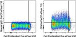
CD49a (Integrin alpha 1) Antibody (46-9490-42) in Flow
Normal human peripheral blood cells were labeled with Cell Proliferation Dye eFluor® 450 (Product # 65-0842-85) then stimulated with Anti-Human CD3 Functional Grade Purified (Product # 16-0037-81), Anti-Human CD28 Functional Grade Purified (Product # 16-0289-81), and Human IL-2 Recombinant Protein (Product # 14-8029-81) for 7 days. Cells were then stained with Mouse IgG1 K Isotype Control PerCP-eFluor® 710 ... View More
 Antibody (46-9490-42) in Flow")
 Antibody (46-9490-42) in Flow")
产品信息
46-9490-42
应用
建议稀释比
已发表文章
产品规格
种属反应
Human
已发表种属
Human
宿主/亚型
Mouse
/ IgG1, kappa
分类
Monoclonal
类型
Antibody
克隆号
TS2/7
偶联物
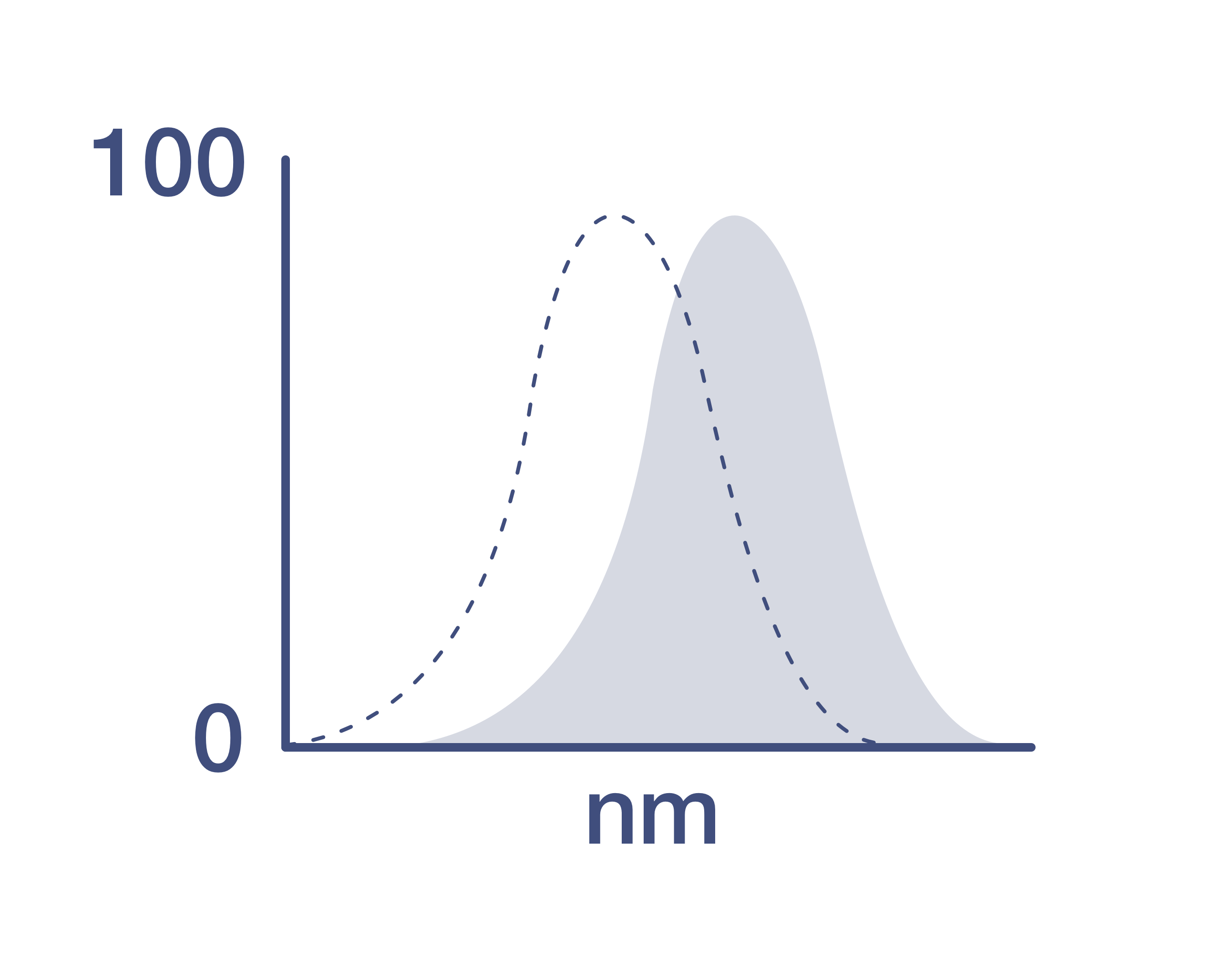
激发/发射光谱
482/708 nm
查看光谱
形式
Liquid
浓度
5 µL/Test
纯化类型
Affinity chromatography
保存液
PBS, pH 7.2, with 0.2% BSA
内含物
0.09% sodium azide
保存条件
4° C, store in dark, DO NOT FREEZE!
运输条件
Ambient (domestic); Wet ice (international)
RRID
AB_2573891
产品详细信息
Description: The monoclonal antibody TS2/7 reacts with human CD49a, also known as integrin alpha 1. CD49a is always associated with CD29 (integrin beta 1) to form a type I transmembrane protein called very late antigen-1 (VLA1). This heterodimer is expressed by endothelial cells, activated T cells, monocytes, a subset of NK cells, a subset of memory T cells, smooth muscle cells, and mesenchymal stem cells. CD49a binds to collagen IV strongly, but also binds to collagen I, laminin, and semaphorin7A. CD49a is involved in cell survival, proliferation, homing, and inflammatory responses.
Applications Reported: This TS2/7 antibody has been reported for use in flow cytometric analysis.
Applications Tested: This TS2/7 antibody has been pre-titrated and tested by flow cytometric analysis of stimulated normal human peripheral blood cells. This can be used at 5 µL (0.125 µg) per test. A test is defined as the amount (µg) of antibody that will stain a cell sample in a final volume of 100 µL. Cell number should be determined empirically but can range from 10^5 to 10^8 cells/test.
PerCP-eFluor® 710 emits at 710 nm and is excited with the blue laser (488 nm); it can be used in place of PerCP-Cyanine5.5. We recommend using a 710/50 bandpass filter, however, the 695/40 bandpass filter is an acceptable alternative. Please make sure that your instrument is capable of detecting this fluorochrome.
Light sensitivity: This tandem dye is sensitive to photo-induced oxidation. Please protect this vial and stained samples from light.
Fixation: Samples can be stored in IC Fixation Buffer (Product # 00-8222) (100 µL of cell sample + 100 µL of IC Fixation Buffer) or 1-step Fix/Lyse Solution (Product # 00-5333) for up to 3 days in the dark at 4°C with minimal impact on brightness and FRET efficiency/compensation. Some generalizations regarding fluorophore performance after fixation can be made, but clone specific performance should be determined empirically.
Excitation: 488 nm; Emission: 710 nm; Laser: Blue Laser.
Filtration: 0.2 µm post-manufacturing filtered.
靶标信息
Integrin alpha 1 (ITGA1) chain associates with the beta 1 (ITGB1) chain to form a heterodimer that functions as a dual laminin/collagen receptor in neural cells and hematopoietic cells. ITGA1 has a 206-amino acid I domain in its N-terminal half, followed by 3 divalent cation-binding sites and a C-terminal transmembrane domain with a short cytoplasmic tail. It also has 28 potential N-glycosylation sites. Human ITGA1 was expressed in a mouse fibroblast cell line as a 180-kD protein. ITGA1 is involved in the early remodeling of osteoarthritic cartilage and plays an essential role in the regulation of mesenchymal stem cell proliferation and cartilage production. It also plays an essential role in the regulation of MSC proliferation and cartilage production.
仅用于科研。不用于诊断过程。未经明确授权不得转售。
How to use the Panel Builder
Watch the video to learn how to use the Invitrogen Flow Cytometry Panel Builder to build your next flow cytometry panel in 5 easy steps.
生物信息学
蛋白别名: CD49 antigen-like family member A; CD49a; integrin alpha 1; Integrin alpha-1; integrin, alpha 1; Laminin and collagen receptor; very late activation protein 1; VLA-1
基因别名: CD49a; ITGA1; VLA1
UniProt ID: (Human) P56199
Entrez Gene ID: (Human) 3672
activation of MAPK activity
muscle contraction
cell-matrix adhesion
integrin-mediated signaling pathway
negative regulation of cell proliferation
extracellular matrix organization
neutrophil chemotaxis
positive regulation of phosphoprotein phosphatase activity
negative regulation of epidermal growth factor receptor signaling pathway
vasodilation
positive regulation of neuron apoptotic process
cellular extravasation
neuron projection morphogenesis

