Search Thermo Fisher Scientific
Disclaimer

Clicking the images or links will redirect you to a website hosted by BenchSci that provides third-party scientific content. Neither the content nor the BenchSci technology and processes for selection have been evaluated by us; we are providing them as-is and without warranty of any kind, including for use or application of the Thermo Fisher Scientific products presented.
 Antibody in Flow Cytometry (Flow)")
图: 1 / 26
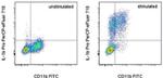
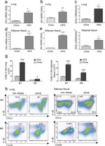
IL-1 beta (Pro-form) Antibody (46-7114-82) in Flow
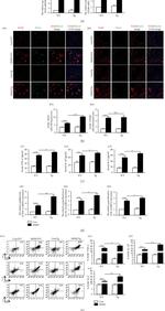
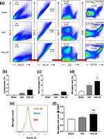
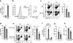
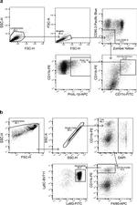
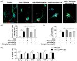
Mouse thioglycolate-elicited peritoneal macrophages were unstimulated (left) or stimulated for 5 hours with LPS (right) in the presence of Protein Transport Inhibitor Cocktail (Product # 00-4980-03). Cells were then fixed and permeabilized with the Intracellular Fixation & Permeabilization Buffer Set (88-8824) then intracellularly stained with h Anti-Mouse CD11b FITC (Product # 11-0112-41) and 0.125 µg of A... View More
 Antibody (46-7114-82) in Flow")
 Antibody (46-7114-82) in Flow")
 Antibody (46-7114-82) in Flow")
 Antibody (46-7114-82) in Flow")
 Antibody (46-7114-82) in Flow")
 Antibody (46-7114-82) in Flow")
 Antibody (46-7114-82) in Flow")
 Antibody (46-7114-82) in Flow")
 Antibody (46-7114-82) in Flow")
 Antibody (46-7114-82) in Flow")
 Antibody (46-7114-82) in Flow")
 Antibody (46-7114-82) in Flow")
 Antibody (46-7114-82) in Flow")
 Antibody (46-7114-82) in Flow")
 Antibody (46-7114-82) in Flow")
 Antibody (46-7114-82) in Flow")
 Antibody (46-7114-82) in Flow")
 Antibody (46-7114-82) in Flow")
 Antibody (46-7114-82) in Flow")
 Antibody (46-7114-82) in Flow")
 Antibody (46-7114-82) in Flow")
 Antibody (46-7114-82) in Flow")
 Antibody (46-7114-82) in Flow")
 Antibody (46-7114-82) in Flow")
 Antibody (46-7114-82) in Flow")
 Antibody (46-7114-82) in Flow")
产品信息
46-7114-82
应用
建议稀释比
已发表文章
产品规格
种属反应
Mouse
已发表种属
Human,
Mouse
宿主/亚型
Rat
/ IgG1, kappa
分类
Monoclonal
类型
Antibody
克隆号
NJTEN3
偶联物
激发/发射光谱
482/708 nm
查看光谱
形式
Liquid
浓度
0.2 mg/mL
纯化类型
Affinity chromatography
保存液
PBS, pH 7.2
内含物
0.09% sodium azide
保存条件
4° C, store in dark, DO NOT FREEZE!
运输条件
Ambient (domestic); Wet ice (international)
RRID
AB_2573835
产品详细信息
Description: This NJTEN3 monoclonal antibody reacts with the pro-form of mouse IL-1 beta, which is a proinflammatory cytokine expressed by monocytes, macrophages, and dendritic cells. It is synthesized in response to inflammatory stimuli as a 31 kDa inactive pro-form that accumulates in the cytosol. Cleavage of pro-IL-1 beta into the active 17 kDa protein requires the activation of inflammasomes, which are multi-protein complexes that respond to pathogens, stress conditions, and other danger signals. Inflammasome activation triggers the processing of the caspase-1 precursor into its active form, which in turn cleaves pro-IL-1 beta. IL-1 beta lacks a signal sequence peptide for classical ER/Golgi pathway and is instead secreted alongside caspase-1 via an alternate and incompletely understood mechanism. IL-1 beta signals via the IL-1RI, which is shared with IL-1 alpha. These cytokines play important roles in innate host defense by triggering the production of other proinflammatory cytokines in target cells and initiating acute-phase responses. Their activity can be moderated by IL-1 Receptor Antagonist (IL-1RA), a protein produced by many cell types that blocks receptor binding through competitive inhibition. Elevated levels of IL-1 beta have been associated with many chronic inflammatory conditions, giving IL-RA or IL-1 beta neutralizing antibodies potential therapeutical value.The NJTEN3 antibody recognizes only the pro-form of mouse IL-1 beta and does not see the active (cleaved) form.
Applications Reported: This NJTEN3 antibody has been reported for use in intracellular staining followed by flow cytometric analysis.
Applications Tested: This NJTEN3 antibody has been tested by intracellular staining and flow cytometric analysis of stimulated mouse thioglycolate-elicited peritoneal macrophages using the Intracellular Fixation & Permeabilization Buffer Set (Product # 88-8824-00) and protocol. Please refer to Best Protocols: Protocol A: Two step protocol for (cytoplasmic) intracellular proteins. This can be used at less than or equal to 0.25 µg per test. A test is defined as the amount (µg) of antibody that will stain a cell sample in a final volume of 100 µL. Cell number should be determined empirically but can range from 10^5 to 10^8 cells/test. It is recommended that the antibody be carefully titrated for optimal performance in the assay of interest.
PerCP-eFluor® 710 emits at 710 nm and is excited with the blue laser (488 nm); it can be used in place of PerCP-Cyanine5.5. We recommend using a 710/50 bandpass filter, however, the 695/40 bandpass filter is an acceptable alternative. Please make sure that your instrument is capable of detecting this fluorochrome.
Fixation: Samples can be stored in IC Fixation Buffer (Product # 00-822-49) (100 µL of cell sample + 100 µL of IC Fixation Buffer) or 1-step Fix/Lyse Solution (Product # 00-5333-54) for up to 3 days in the dark at 4°C with minimal impact on brightness and FRET efficiency/compensation. Some generalizations regarding fluorophore performance after fixation can be made, but clone specific performance should be determined empirically.
Excitation: 488 nm; Emission: 710 nm; Laser: Blue Laser.
Filtration: 0.2 µm post-manufacturing filtered.
靶标信息
Interleukin-1 beta (IL-1 beta) is a proinflammatory cytokine expressed by monocytes, macrophages, and dendritic cells. It is synthesized in response to inflammatory stimuli as a 31 kDa inactive pro-form that accumulates in the cytosol. Cleavage of pro-IL-1 beta into the active 17 kDa protein requires the activation of inflammasomes, which are multi-protein complexes that respond to pathogens, stress conditions, and other danger signals. Inflammasome activation triggers the processing of the caspase-1 precursor into its active form, which in turn cleaves pro-IL-1 beta. IL-1 beta lacks a signal sequence peptide for classical ER/Golgi pathway and is instead secreted alongside caspase-1 via an alternate and incompletely understood mechanism. Although IL-1 beta is most often secreted in its active form, secretion of the uncleaved protein may be detectable under some biological conditions.IL-1 beta signals through two receptors, IL-1RI and IL-1RII, both of which are shared with IL-1 alpha. These cytokines play important roles in innate host defense by triggering the production of other proinflammatory cytokines in target cells and initiating acute-phase responses. Their activity can be moderated by IL-1 Receptor Antagonist (IL-1RA), a protein produced by many cell types that blocks receptor binding through competitive inhibition. Elevated levels of IL-1 beta have been associated with many chronic inflammatory conditions, giving IL-RA or IL-1 beta neutralizing antibodies potential therapeutical value.
仅用于科研。不用于诊断过程。未经明确授权不得转售。
How to use the Panel Builder
Watch the video to learn how to use the Invitrogen Flow Cytometry Panel Builder to build your next flow cytometry panel in 5 easy steps.
生物信息学
蛋白别名: il 1b; IL 1β; IL-1 beta; IL1β; ILN; Interleukin; Interleukin-1 beta; Interleukin-1b; Interleukin1 beta
基因别名: Il-1b; IL-1beta; Il1b
UniProt ID: (Mouse) P10749
Entrez Gene ID: (Mouse) 16176
negative regulation of transcription from RNA polymerase II promoter
MAPK cascade
activation of MAPK activity
fever generation
positive regulation of protein phosphorylation
inflammatory response
immune response
positive regulation of cytosolic calcium ion concentration
aging
memory
negative regulation of cell proliferation
response to carbohydrate
positive regulation of vascular endothelial growth factor production
positive regulation of gene expression
negative regulation of gene expression
negative regulation of glucose transport
positive regulation of cell death
negative regulation of glutamate secretion
cytokine-mediated signaling pathway
hyaluronan biosynthetic process
neutrophil chemotaxis
sequestering of triglyceride
positive regulation of fever generation
lipopolysaccharide-mediated signaling pathway
positive regulation of prostaglandin secretion
response to lipopolysaccharide
interleukin-1 beta production
positive regulation of granulocyte macrophage colony-stimulating factor production
positive regulation of interferon-gamma production
positive regulation of interleukin-6 production
positive regulation of interleukin-8 production
positive regulation of stress-activated MAPK cascade
positive regulation of immature T cell proliferation in thymus
response to ATP
positive regulation of heterotypic cell-cell adhesion
social behavior
ectopic germ cell programmed cell death
positive regulation of myosin light chain kinase activity
cellular response to drug
positive regulation of T cell proliferation
positive regulation of NF-kappaB import into nucleus
positive regulation of apoptotic process
regulation of I-kappaB kinase/NF-kappaB signaling
positive regulation of I-kappaB kinase/NF-kappaB signaling
negative regulation of MAP kinase activity
protein kinase B signaling
positive regulation of JUN kinase activity
positive regulation of chemokine biosynthetic process
positive regulation of interleukin-2 biosynthetic process
positive regulation of interleukin-6 biosynthetic process
positive regulation of nitric oxide biosynthetic process
negative regulation of neuron differentiation
positive regulation of glial cell differentiation
positive regulation of angiogenesis
negative regulation of lipid metabolic process
positive regulation of mitotic nuclear division
positive regulation of transcription, DNA-templated
positive regulation of transcription from RNA polymerase II promoter
positive regulation of JNK cascade
negative regulation of insulin receptor signaling pathway
positive regulation of astrocyte differentiation
positive regulation of phagocytosis
negative regulation of neurogenesis
regulation of insulin secretion
leukocyte migration
negative regulation of lipid catabolic process
positive regulation of lipid catabolic process
positive regulation of membrane protein ectodomain proteolysis
positive regulation of sequence-specific DNA binding transcription factor activity
positive regulation of NF-kappaB transcription factor activity
positive regulation of cell division
positive regulation of calcidiol 1-monooxygenase activity
negative regulation of adiponectin secretion
positive regulation of ERK1 and ERK2 cascade
monocyte aggregation
cellular response to organic substance
cellular response to organic cyclic compound
positive regulation of monocyte chemotactic protein-1 production
positive regulation of neutrophil chemotaxis
extrinsic apoptotic signaling pathway in absence of ligand
negative regulation of branching morphogenesis of a nerve
negative regulation of neural precursor cell proliferation
negative regulation of extrinsic apoptotic signaling pathway in absence of ligand

