Search Thermo Fisher Scientific
Disclaimer

Clicking the images or links will redirect you to a website hosted by BenchSci that provides third-party scientific content. Neither the content nor the BenchSci technology and processes for selection have been evaluated by us; we are providing them as-is and without warranty of any kind, including for use or application of the Thermo Fisher Scientific products presented.
")
图: 1 / 1
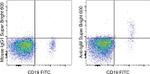
IgM Antibody (63-9998-42) in Flow
Staining of normal human peripheral blood cells with Anti-Human CD19 FITC (Product # 11-0199-42) and Mouse IgG1 K Isotype Control Super Bright 600 (Product # 63-4714-82) (left) or Anti-Human IgM Super Bright 600 (right). Cells in the lymphocyte gate were used for analysis.
 in Flow")
产品信息
63-9998-42
产品规格
种属反应
Human
宿主/亚型
Mouse
/ IgG1, kappa
分类
Monoclonal
类型
Antibody
克隆号
SA-DA4
偶联物
激发/发射光谱
414/601 nm
查看光谱
形式
Liquid
浓度
5 µL/Test
纯化类型
Affinity chromatography
保存液
PBS, pH 7.2, with BSA
内含物
0.09% sodium azide
保存条件
4° C, store in dark, DO NOT FREEZE!
运输条件
Ambient (domestic); Wet ice (international)
RRID
AB_2662359
产品详细信息
Description: The SA-DA4 monoclonal antibody reacts with the mu heavy chain of human IgM. It does not react with other classes of human immunoglobulin including IgD, IgG, or IgA. IgM is expressed intracellularly, during early stages of B lymphopoiesis, and then on the surface of mature B cells.
Applications Reported: This SA-DA4 antibody has been reported for use in flow cytometric analysis.
Applications Tested: This SA-DA4 antibody has been pre-titrated and tested by flow cytometric analysis of normal human peripheral blood cells. This can be used at 5 µL (0.125 µg) per test. A test is defined as the amount (µg) of antibody that will stain a cell sample in a final volume of 100 µL. Cell number should be determined empirically but can range from 105 to 108 cells/test.
Super Bright 600 is a tandem dye that can be excited with the violet laser line (405 nm) and emits at 600 nm. We recommend using a 610/20 bandpass filter. Please make sure that your instrument is capable of detecting this fluorochrome.
When using two or more Super Bright dye-conjugated antibodies in a staining panel, it is recommended to use Super Bright Complete Staining Buffer (Product # SB-4401) to minimize any non-specific polymer interactions. Please refer to the datasheet for Super Bright Staining Buffer for more information.
Light sensitivity: This tandem dye is sensitive to photo-induced oxidation. Protect this vial and stained samples from light.
Fixation: Samples can be stored in IC Fixation Buffer (Product # 00-8222) (100 µL of cell sample + 100 µL of IC Fixation Buffer) or 1-step Fix/Lyse Solution (Product # 00-5333) for up to 3 days in the dark at 2-8°C with minimal impact on brightness and FRET efficiency/compensation. Some generalizations regarding fluorochrome performance after fixation can be made, but clone specific performance should be determined empirically.
Excitation: 405 nm; Emission: 600 nm; Laser: Violet Laser
Super Bright Polymer Dyes are sold under license from Becton, Dickinson and Company.
靶标信息
IgM is expressed intracellularly, during early stages of B lymphopoiesis, and then on the surface of more mature B cells in the bone marrow and peripheral B cells.
仅用于科研。不用于诊断过程。未经明确授权不得转售。
How to use the Panel Builder
Watch the video to learn how to use the Invitrogen Flow Cytometry Panel Builder to build your next flow cytometry panel in 5 easy steps.
篇参考文献 (0)
您是否在文献中引用过该产品?请点击下方按钮邮件告知我们。
生物信息学
基因别名: AGM1; MU; VH
Entrez Gene ID: (Human) 3507

