Search Thermo Fisher Scientific
Disclaimer

Clicking the images or links will redirect you to a website hosted by BenchSci that provides third-party scientific content. Neither the content nor the BenchSci technology and processes for selection have been evaluated by us; we are providing them as-is and without warranty of any kind, including for use or application of the Thermo Fisher Scientific products presented.
")
图: 1 / 5
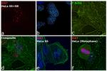
Ki-67 Antibody (606-5698-82) in ICC/IF
Immunofluorescence analysis of Ki-67 Monoclonal Antibody (SolA15), Alexa Fluor™ 660, eBioscience™ was performed using 70% confluent log phase HeLa cells serum starved (SS) for 36 hours followed by serum release (SR) for 6 hours. The cells were fixed with 4% paraformaldehyde for 10 minutes, permeabilized with 0.1% Triton™ X-100 for 15 minutes, and blocked with 2% BSA for 45 minutes at room temperature. The cells were labeled with Ki-67 Monoclonal Antibody (SolA15), Alexa Fluor™ 660, eBioscience™ (Product # 606-5698-82) at 1:100 dilution in ... View More

 in ICC/IF")
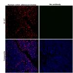
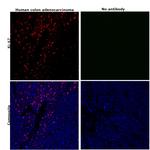
 in IHC (P)")
 in IHC (P)")
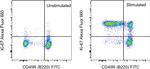
 in Flow")
")
产品信息
606-5698-82
产品规格
种属反应
Dog,
Cynomolgus monkey,
Human,
Mouse,
Non-human primate,
Rat
宿主/亚型
Rat
/ IgG2a, kappa
分类
Monoclonal
类型
Antibody
克隆号
SolA15
偶联物
激发/发射光谱
663/691 nm
查看光谱
形式
Liquid
浓度
0.2 mg/mL
纯化类型
Affinity chromatography
保存液
PBS, pH 7.2
内含物
0.09% sodium azide
保存条件
4° C, store in dark, DO NOT FREEZE!
运输条件
Ambient (domestic); Wet ice (international)
RRID
AB_2896286
产品详细信息
Description: The monoclonal antibody SolA15 recognizes mouse and rat Ki-67, a 300 kDa nuclear protein. Ki-67 is present during all active phases of the cell cycle (G1, S, G2, and mitosis), but is absent from resting cells (G0). Ki-67 is detected within the nucleus during interphase but redistributes to the chromosomes during mitosis. Ki-67 is used as a marker for determining the growth fraction of a given population of cells. In studies of tumor cells, the "Ki-67 labeling index" refers to the number of Ki-67 positive cells within the population and this is used to predict outcome of particular cancer types. Ki-67 has been shown to interact with the DNA-bound protein chromobox protein homolog 3 (CBX3) (heterochromatin).
The SolA15 antibody also recognizes human, non-human primate and canine Ki-67.
Applications Reported: This SolA15 antibody has been reported for use in intracellular staining followed by flow cytometric analysis.
Applications Tested: This SolA15 antibody has been tested by intracellular staining followed by flow cytometric analysis of stimulated mouse splenocytes using the Foxp3/Transcription Factor Staining Buffer Set (Product # 00-5523-00) and protocol. Please refer to "Staining Intracellular Antigens for Flow Cytometry, Protocol B: One step protocol for intracellular (nuclear) proteins" located at Flow Protocols . This may be used at less than or equal to 0.25 µg per test. A test is defined as the amount (µg) of antibody that will stain a cell sample in a final volume of 100 µL. Cell number should be determined empirically but can range from 10^5 to 10^8 cells/test. It is recommended that the antibody be carefully titrated for optimal performance in the assay of interest.
Alexa Fluor 660 emits at 690 nm and is intended for use on spectral cytometers where it may be multiplexed with APC, Alexa Fluor 647 and Alexa Fluor 700.
Excitation: 662 nm; Emission: 690 nm; Laser: Red Laser.
靶标信息
Ki-67 is a nuclear protein that is expressed during various stages in the cell cycle, particularly during late G1, S, G2, and M phases. The protein has a forkhead associated domain (FHA) through which it associates with euchromatin at the perichromosomal layer, the centromeric heterochromatin, and the nucleolus. Ki-67 is shown to have a cell cycle dependent topographical distribution with perinucleolar expression at G1, expression in the nuclear matrix at G2, and expression on the chromosomes during M phase. Ki-67 is commonly used as a proliferation marker because it is not detected in G0 cells, but increases steadily from G1 through mitosis. Ki-67 antibodies are useful in establishing the cell growing fraction in neoplasms. In neoplastic tissues, the prognostic value is comparable to the tritiated thymidine-labelling index. The correlation between low Ki-67 index and histologically low-grade tumors is strong. Ki-67 is routinely used as a neuronal marker of cell cycling and proliferation.
仅用于科研。不用于诊断过程。未经明确授权不得转售。
How to use the Panel Builder
Watch the video to learn how to use the Invitrogen Flow Cytometry Panel Builder to build your next flow cytometry panel in 5 easy steps.
篇参考文献 (0)
您是否在文献中引用过该产品?请点击下方按钮邮件告知我们。
生物信息学
蛋白别名: Antigen identified by monoclonal antibody Ki-67; Antigen identified by monoclonal antibody Ki-67 homolog; Antigen KI-67; Antigen KI-67 homolog; Proliferation marker protein Ki-67; proliferation-related Ki-67 antigen; protein phosphatase 1, regulatory subunit 105; RP11-380J17.2; unnamed protein product
基因别名: D630048A14Rik; Ki-67; Ki67; KIA; MIB-; MIB-1; MKI67; PPP1R105
UniProt ID: (Human) P46013, (Mouse) E9PVX6
Entrez Gene ID: (Dog) 100686578, (Human) 4288, (Cynomolgus monkey) 102135895, (Rat) 291234, (Mouse) 17345

