Search Thermo Fisher Scientific
Disclaimer

Clicking the images or links will redirect you to a website hosted by BenchSci that provides third-party scientific content. Neither the content nor the BenchSci technology and processes for selection have been evaluated by us; we are providing them as-is and without warranty of any kind, including for use or application of the Thermo Fisher Scientific products presented.
")
图: 1 / 2
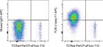
TCR V beta F1 Antibody (17-5766-42) in Flow
Human PBMC were stained with CD3 Monoclonal Antibody, PE (Product # 12-0038-42) and TCR gamma/delta Monoclonal Antibody, PerCP-eFluor 710 (Product # 46-9959-42). The cells were then fixed and permeabilized using the Intracellular Fixation & Permeabilization Buffer Set (Product # 88-8824-00) and protocol, and stained with Mouse IgG1 kappa Isotype Control, APC (Product # 17-4714-42) (left) or TCR beta F1 Monoclonal Antibody, PE-Cyanine 7 (right). Viable CD3 positive lymphocytes were used for analysis.
 in Flow")
")
产品信息
17-5766-42
产品规格
种属反应
Human
宿主/亚型
Mouse
/ IgG1, kappa
分类
Monoclonal
类型
Antibody
克隆号
8A3
抗原
Human TCR beta chain constant region
偶联物
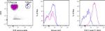
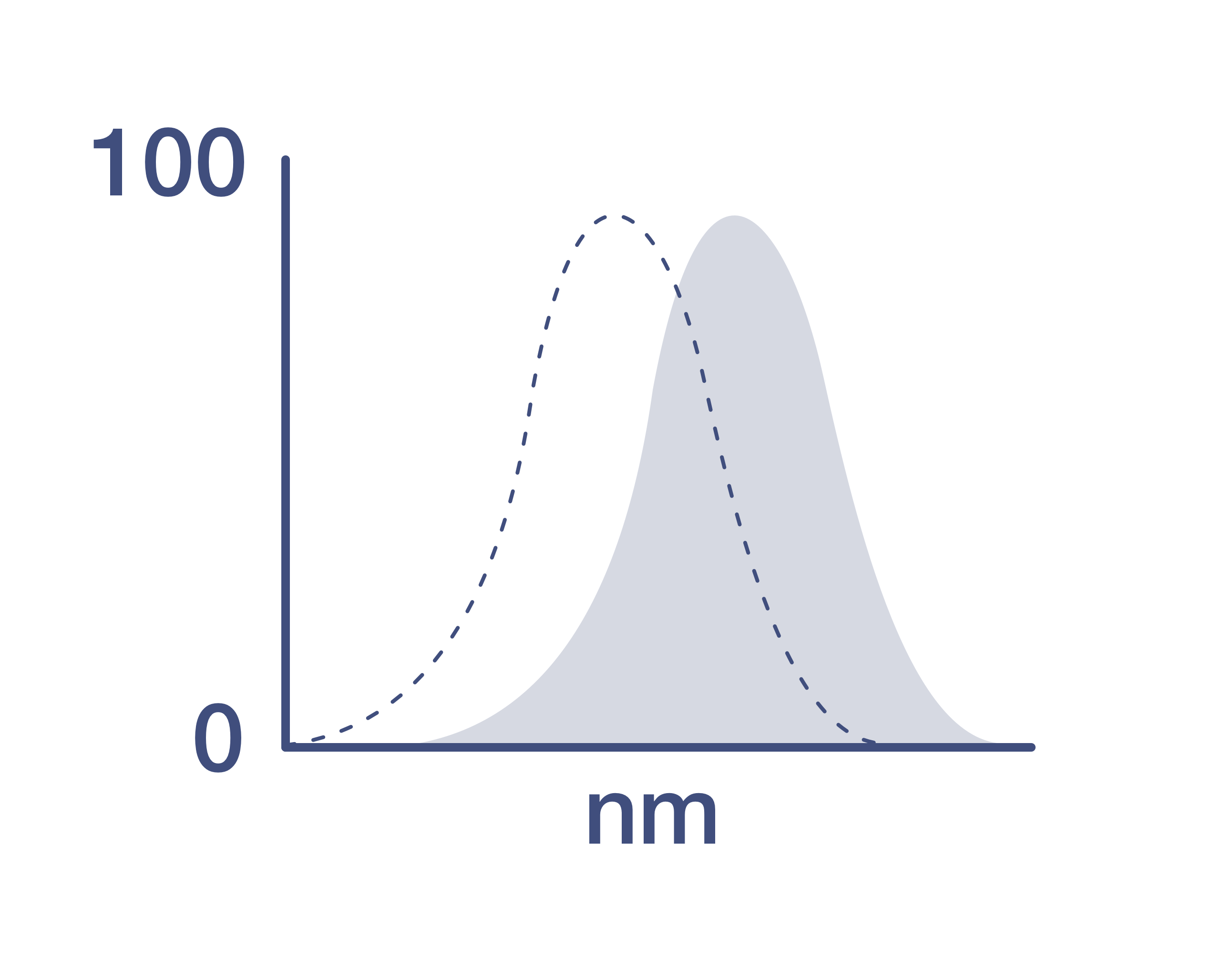
激发/发射光谱
651/660 nm
查看光谱
形式
Liquid
浓度
5 µL/Test
纯化类型
Affinity chromatography
保存液
PBS, pH 7.2, with 0.2% BSA
内含物
0.09% sodium azide
保存条件
4° C, store in dark, DO NOT FREEZE!
运输条件
Ambient (domestic); Wet ice (international)
RRID
AB_2744710
产品详细信息
Description: This 8A3 monoclonal antibody recognizes human TCR beta F1, a common framework determinant on the beta-subunit of the T cell receptor. Expression of TCR beta F1 has been described as a potential diagnostic marker indicating a favorable outcome for tumor patients with not otherwise specified Nodal Peripheral T-cell Lymphoma (PTCL-NOS). Furthermore, a lack of surface and cytoplasmic TCR beta F1 was also found in small cell variant of T-cell prolymphocytic Leukemia. This antibody will not stain the native form of the TCR and will require fixing and permeabilizing cells.
Applications Reported: This 8A3 antibody has been reported for use in flow cytometric analysis, immunofluorescent staining of frozen tissue sections, and immunocytochemistry.
Applications Tested: This 8A3 antibody has been pre-diluted and tested by flow cytometric analysis of normal human peripheral blood cells. This may be used at 5 µL (0.125 µg) per test. A test is defined as the amount (µg) of antibody that will stain a cell sample in a final volume of 100 µL. Cell number should be determined empirically but can range from 10^5 to 10^8 cells/test.
Excitation: 633-647 nm; Emission: 660 nm; Laser: Red Laser
靶标信息
The T Cell Receptor alpha beta (TCRab) is a heterodimeric membrane receptor that consists of the highly variable alpha and beta chains expressed as part of a complex with the invariant CD3 proteins. TCRab participates in the activation of T-cells in response to an antigen presented in the context of MHC. Engagement of the TCRab initiates signaling cascades that ultimately result in cellular proliferation, differentiation, cytokine production, and/or activation-induced cell death. These signaling cascades regulate T-cell development, homeostasis, activation, acquisition of effector functions and apoptosis. Cells expressing TCRab constitute the vast majority of T cells in both human and mouse.
仅用于科研。不用于诊断过程。未经明确授权不得转售。
How to use the Panel Builder
Watch the video to learn how to use the Invitrogen Flow Cytometry Panel Builder to build your next flow cytometry panel in 5 easy steps.
篇参考文献 (0)
您是否在文献中引用过该产品?请点击下方按钮邮件告知我们。

