Search Thermo Fisher Scientific
Disclaimer

Clicking the images or links will redirect you to a website hosted by BenchSci that provides third-party scientific content. Neither the content nor the BenchSci technology and processes for selection have been evaluated by us; we are providing them as-is and without warranty of any kind, including for use or application of the Thermo Fisher Scientific products presented.
")
图: 1 / 4
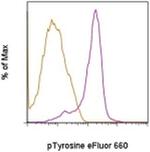
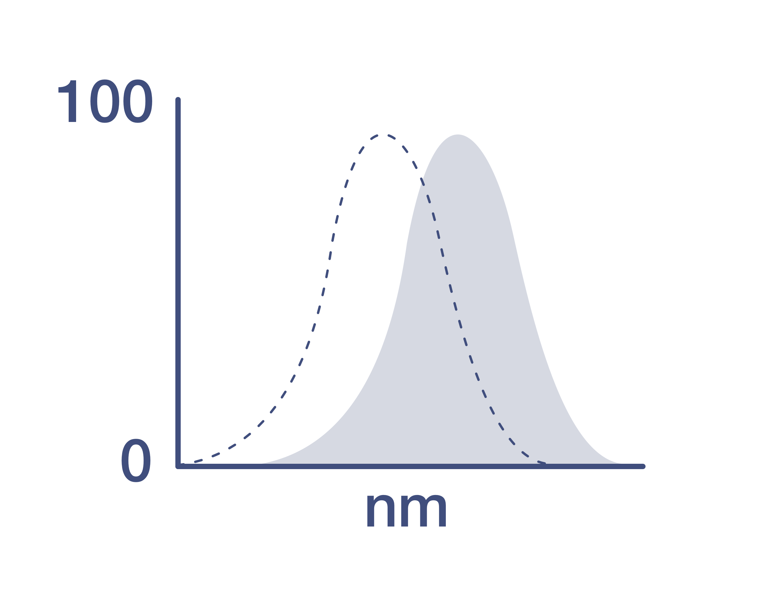
phospho-Tyrosine Antibody (50-5001-42) in Flow
Intracellular staining of normal human peripheral blood cells left untreated (orange histogram) or treated with hydrogen-peroxide-activated sodium pervanadate (purple histogram) and stained with 0.06 µg of Anti-Human/Mouse phospho-Tyrosine eFluor® 660 (purple histogram) using the IC Fixation & Permeabilization Buffer Set (Product # 88-8824-00). Cells in the lymphocyte gate were used for analysis.
 in Flow")
 in ICC/IF")
 in ICC/IF")
 in Flow")
产品信息
50-5001-42
应用
建议稀释比
已发表文章
产品规格
种属反应
Chemical
已发表种属
Not Applicable
宿主/亚型
Mouse
/ IgG2b, kappa
分类
Monoclonal
类型
Antibody
克隆号
pY20
偶联物
激发/发射光谱
651/668 nm
查看光谱
形式
Liquid
浓度
5 µL/Test
纯化类型
Affinity chromatography
保存液
PBS, pH 7.2, with 0.2% BSA
内含物
0.09% sodium azide
保存条件
4° C, store in dark, DO NOT FREEZE!
运输条件
Ambient (domestic); Wet ice (international)
RRID
AB_2574231
产品详细信息
Description: The pY20 monoclonal antibody recognizes phosphorylated tyrosine residues (p-Tyr) on proteins. Numerous intracellular signaling cascades are propagated via phosphorylation of specific tyrosine on signaling proteins. The detection of p-Tyr residues is valuable for the characterization and purification of phosphorylated proteins and the biochemical pathways that they are involved in.
Applications Reported: This pY20 antibody has been reported for use in intracellular staining followed by flow cytometric analysis.
Applications Tested: This pY20 antibody has been pre-titrated and tested by intracellular staining followed by flow cytometric analysis of normal human peripheral blood cells. This can be used at 5 µL (0.06 µg) per test. A test is defined as the amount (µg) of antibody that will stain a cell sample in a final volume of 100 µL. Cell number should be determined empirically but can range from 10^5 to 10^8 cells/test.
Staining Protocol: All protocols work well for this monoclonal antibody. Use of Protocol A: Two-step protocol: intracellular (cytoplasmic) proteins allows for the greatest flexibility for detection of surface and intracellular (cytoplasmic) proteins. Use of Protocol B: One-step protocol: intracellular (nuclear) proteins is recommended for staining of transcription factors in conjunction with surface and phosphorylated intracellular (cytoplasmic) protein(s). Protocol C: Two-step protocol: Fixation/Methanol allows for the greatest discrimination of phospho-specific signaling between unstimulated and stimulated samples, but with limitations on the ability to stain specific surface proteins (refer to "Clone Performance Following Fixation/Permeabilization" located in the Best Protocols Section under the Resources tab online). All Protocols can be found in the Flow Cytometry Protocols: "Staining Intracellular Antigens for Flow Cytometry Protocol" located in the Best Protocols Section under the Resources tab online.
Excitation: 633-647 nm; Emission: 668 nm; Laser: Red Laser.
Filtration: 0.2 µm post-manufacturing filtered.
靶标信息
The role of tyrosine phosphorylation in transduction of the mitogenic signal from transmembrane receptors and in transformation by oncogene tyrosine kinases has been the subject of intense investigation for several years. While the phosphorylation of specific tyrosine residues has been shown to be a primary mechanism of signal transduction during normal mitogenesis, cell cycle progression and oncogenic transformation, its role in other areas such as differentiation and gap junction communication, is a matter of active and ongoing research. Antibodies that specifically recognize phosphorylated tyrosine residues have proved to be invaluable to the study of tyrosine -phosphorylated proteins and the biochemical pathways in which they function. The fluorescein (FITC) conjugate of clone PY20 anti-phosphotyrosine is especially useful for the detection of these P-Tyr proteins in immunohistochemical and immunocytochemical protocols in situations wherein the use of a secondary antibody would complicate detection of the protein(s) of interest.
仅用于科研。不用于诊断过程。未经明确授权不得转售。
How to use the Panel Builder
Watch the video to learn how to use the Invitrogen Flow Cytometry Panel Builder to build your next flow cytometry panel in 5 easy steps.
生物信息学
蛋白别名: Phosphotyrosine; pTyr; pY

